Health & New Tech
Women's Health
Post-Doctoral Fellowships
United States
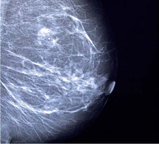
Enhanced molecular Breast Cancer imaging : 1 mm resolution positron emission tomography
My research focuses on developing novel, high resolution imaging instrumentation for organ or patient specific Positron Emission Tomography (PET), a non-invasive, in-vivo molecular imaging technology. Currently efforts are focused on constructing a high-performance, low cost, portable camera dedicated to the detection and diagnosis of breast cancer.
3D imaging techniques for cancer management
Virginia Spanoudaki is currently holding a post-doctoral grant at the prestigious Stanford University (California, USA), within the Molecular imaging Program (MIPS). Apart from research, she is also involved in teaching and other educational activities.
“I currently work on developing a new instrument for tomographic medical imaging that, if successful, will substantially enhance image quality and accuracy with a focus of benefiting breast cancer management. The technique improves our ability to characterise the biology of the disease well before structural changes result.
Working on developing technologies to help the management of disease such as breast cancer can be challenging, in many ways, from both scientific and human points of view. For example, currently there are difficulties associated with the detection, diagnosis and staging of breast cancer.
Mammography is accepted as the best means to screen for non-palpable breast cancer; however the current technologies have a very high rate of false-positive detection (around 80%)! There are every year in the US more than 600, 000 unnecessary biopsies, which costs between $ 1,000 to $ 3,000 per procedure.
By combining physics, engineering and biology, the purpose is for us to use physical principles to image human diseases. Medical imaging is a very active field of research in many centers around the world, with instrumentation being only one, but extremely important, part of it. Our lab is specialised in developing novel detector technologies for “Positron Emission Tomography” (PET). PET is a non-invasive, molecular imaging technology that has shown promises of a more accurate identification of cancer than other imaging modalities due to its unique ability to sense and visualise increased biochemical changes in malignant tissues, compared to healthy ones, before structural changes occur! PET technology is used for diagnosis and staging of disease, as well as for monitoring of treatment. It has a widespread application in oncology, neurology and cardiology.
The beauty of my field of research is that it is multidisciplinary: Professor Levin’s lab, the MIIL, where I am currently a post-doctoral fellow, is composed of physicists, electrical, computer, mechanical, material and biomedical engineers. Furthermore, in our programme we also interact with biologists, chemists and physicians. In this way we can all benefit from the expertise of each other and learn from everyone’s scientific experience. In my opinion this is what makes a multidisciplinary field, such as molecular imaging, so attractive.
As a scientist, I announce the results of my research through journal publications or participation in conferences where I can present some of my results. These are common ways to exchange ideas with scientists worldwide and build a scientific network. One such conference is the IEEE Medical Imaging Conference (see www.nss-mic.org).”
To add or modify information on this page, please contact us at the following address: community.research@axa.com

Virginia
SPANOUDAKI
Institution
Stanford University
Country
United States
Nationality
American
Related articles
Women's Health
AXA Chair
Spain
Understanding Menopause's Effect on Women's Cardiovascular Risk
Cardiovascular disease (CVD) is the leading cause of mortality for women worldwide, yet women remain understudied. Atherosclerosis, the main pathology underlying... Read more
.thumbnail.jpg)
Inés
PINEDA-TORRA
University of Seville
Mental Health & Neurology
Women's Health
Pandemics & Infectious Diseases
AXA Award
Spain
The Effects of the COVID-19 Pandemic on the Mental Health of Mothers and Newborns
Early results are expected in less than 12 months and will help bridge a critical gap in research. Indeed, “in... Read more

Maria
FORASTER
ISGlobal Barcelona Institute for Global Health
Health & New Tech
Pandemics & Infectious Diseases
Artificial Intelligence
AXA Award
Spain
Early Prognosis of COVID-19 Infections Via Machine Learning
Research has already shown that certain health data, such as age and past medical history (PMH), have a strong correlation... Read more

Santiago
MAZUELAS